Common Symptoms of Keratoconus
Keratoconus symptoms often begin gradually and worsen over time. Early signs may resemble ordinary refractive error but become more distinct as the cornea steepens.
- Blurred or distorted vision that does not fully correct with glasses
- Increased sensitivity to light and glare
- Halos or streaking around lights, especially at night
- Frequent changes in glasses or contact lens prescriptions
- Difficulty with night driving or reading fine print
- Double vision or ghost images in one eye
Why Glasses Often Do Not Work for Keratoconus
Glasses correct vision by adjusting how light enters the eye, but they rely on the cornea having a relatively uniform shape. In keratoconus, the cornea becomes irregularly curved, scattering light in multiple directions. Glasses cannot compensate for this type of distortion, which is why patients often report that their vision remains blurry even with an updated prescription. As the condition progresses, specialty contact lenses are typically required to provide a smooth optical surface over the irregular cornea.
Is Keratoconus a Progressive Condition?
Yes, keratoconus is typically a progressive condition. It most commonly advances during the teenage years and twenties, though the rate of progression varies among individuals. In many patients, the condition stabilizes by the mid-30s to 40s. Corneal cross-linking is a procedure that can slow or halt progression when performed early. Regular monitoring with corneal imaging is important to detect changes over time and adjust the management plan accordingly.
Can Keratoconus Cause Permanent Vision Loss?
Keratoconus does not typically cause complete blindness, but it can lead to significant and permanent vision impairment if left unmanaged. In advanced cases, corneal scarring may develop and reduce best-corrected visual acuity. In rare instances, a condition called corneal hydrops can cause sudden swelling and further scarring. With appropriate management, including specialty contact lenses and monitoring, most patients maintain functional vision throughout their lives.
What Happens If Keratoconus Is Not Treated
Without management, keratoconus may continue to progress, leading to increasing corneal irregularity and worsening vision. Over time, thinning and steepening of the cornea can result in scarring that further reduces visual clarity. In advanced stages, contact lens fitting becomes more difficult, and corneal transplantation may become the remaining option. Early detection and ongoing monitoring allow for timely interventions that can preserve corneal integrity and visual function.
Contact Lens Options for Keratoconus
Several types of contact lenses are used to manage keratoconus. The appropriate option depends on the severity of the condition and the individual shape of the cornea.
Glasses
Glasses may provide adequate correction in early-stage keratoconus. As the cornea becomes more irregular, glasses are generally unable to correct the resulting distortion and are no longer sufficient as a primary form of vision correction.
Rigid Gas Permeable (RGP) Lenses
RGP lenses are small, rigid lenses that rest directly on the cornea. They create a smooth refracting surface that can improve visual acuity in mild to moderate keratoconus. Some patients find them less comfortable than other options, particularly when the cornea is highly irregular.
Hybrid Lenses
Hybrid lenses combine a rigid center with a soft outer skirt. They offer some of the optical clarity of an RGP lens with improved comfort. They may be appropriate for patients with moderate keratoconus who have difficulty wearing rigid lenses.
Scleral Lenses — Beyond Keratoconus
Scleral lenses are large-diameter rigid gas permeable lenses that vault over the entire cornea and rest on the sclera (the white part of the eye). A fluid reservoir between the lens and cornea creates a smooth optical surface, bypassing corneal irregularity. Scleral lenses are often the preferred option for moderate to advanced keratoconus because they provide consistent visual correction, improved comfort compared to smaller rigid lenses, and protection for the corneal surface.
Scleral lenses are not limited to keratoconus. They are also used to restore vision in patients with other forms of corneal irregularity, including:
- Post-keratoplasty (corneal transplant): After a corneal graft, the new cornea often heals with an irregular surface that glasses cannot correct. Scleral lenses vault over the graft and provide stable, clear vision.
- Post-LASIK ectasia: In rare cases, LASIK can weaken the cornea and cause it to bulge forward, producing irregular astigmatism similar to keratoconus. Scleral lenses are often the most effective non-surgical solution for restoring visual clarity.
- Post-RK (radial keratotomy): Patients who had radial keratotomy in the 1980s and 1990s often experience fluctuating vision and progressive corneal irregularity decades later. Scleral lenses provide a stable optical surface over the RK incision scars.
- Pellucid marginal degeneration: A condition similar to keratoconus where thinning occurs at the lower periphery of the cornea, causing irregular astigmatism that scleral lenses can correct.
Dr. Bonakdar has extensive experience fitting scleral lenses for all of these conditions. Learn more about scleral lenses for post-surgical eyes →
How Keratoconus Is Monitored Over Time
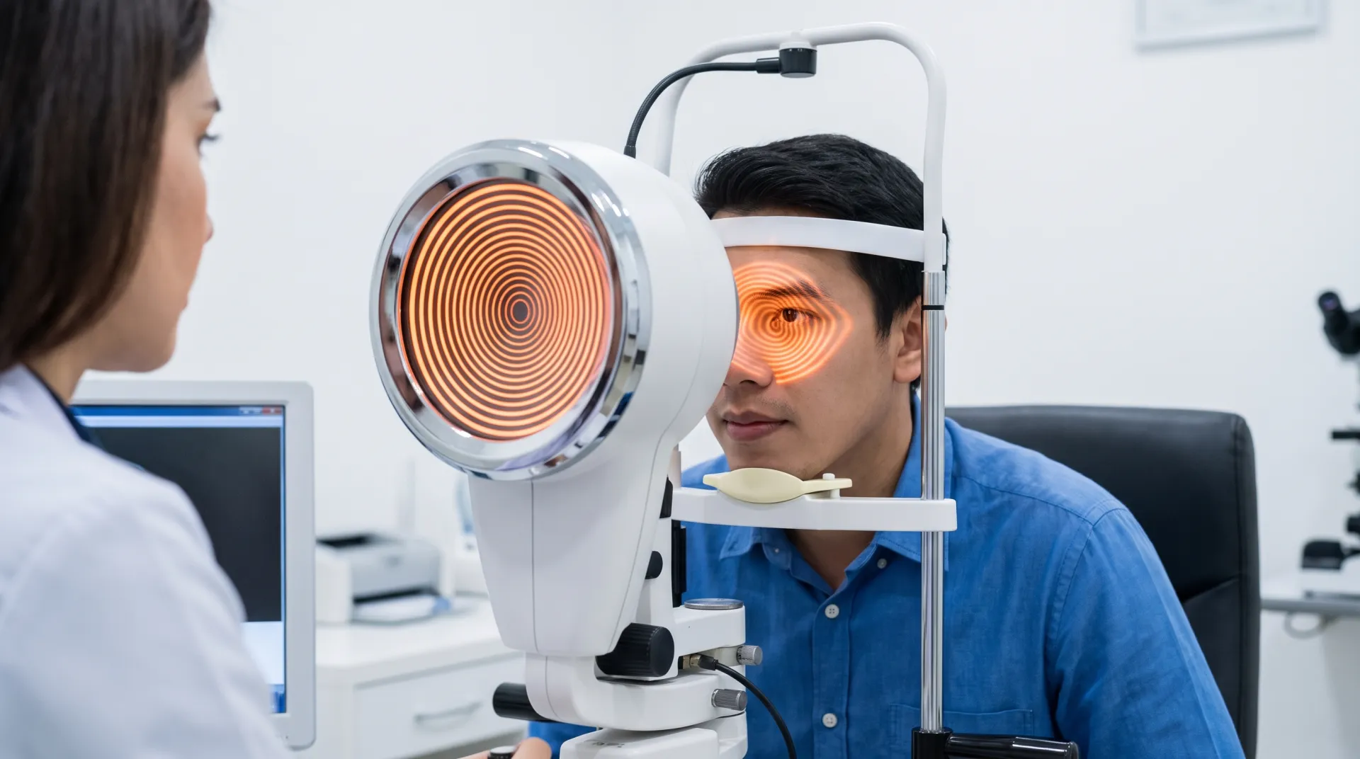
Keratoconus is monitored through regular comprehensive eye examinations that include corneal topography and tomography. These imaging techniques create detailed maps of the corneal surface and thickness, allowing clinicians to detect subtle changes in shape over time. Monitoring frequency depends on the patient's age, rate of progression, and current management approach. Patients who have undergone corneal cross-linking or who wear scleral lenses are typically seen at regular intervals to assess corneal stability and lens fit.